????Amira for Life Sciences??
??????3D/4D+ visualization and analysis software??
Thermo Scientific Amira Software is a powerful, multifaceted 3D/4D+ platform for visualizing, manipulating, and understanding life science research data from many image modalities, including CT, MRI, 3D Microscopy, and other techniques.
With incredible speed and flexibility, Amira Software enables advanced 3D/4D+ bioimaging workflows in research areas ranging from structural and cellular biology, to tissue imaging, neuroscience, preclinical imaging, and bioengineering.
??????

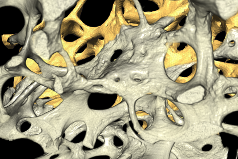
??Image is courtesy of Elsa Vennat, CNRS – Laboratoire MSSMat (France)??
??
??Cell biology??
Understanding living cells and their processes requires analysis of data from various imaging systems and modalities that all use different file formats. Each experimental setup can represent a unique challenge to process the data. Multi-scale dynamic processes require tracking of various sized objects, from diffraction-limited particles to entire cells. There can be a few dozen to tens of thousands objects in each data set. In addition, intracellular processes are supported by sub-cellular structures that also need to be identified and quantified.
Amira Software provides a comprehensive array of tools for the flexible and accurate analysis of time series data of cellular processes. It enables researchers to perform dedicated segmentation workflows on their intra- and intercellular images and apply powerful automated object tracking solution.??
??Preclinical imaging??
????Preclinical image analysis relies on augmenting structural information from micro-CT or MRI systems with functional data from PET, SPECT or optical imaging instrumentation. Analysis of novel drug candidates requires pinpointing the location of dynamic processes acquired with functional modalities in images of the animal’s anatomy from structural modalities over time.??
Amira Software enables the fusion of any type of preclinical image data. Co-registered images can be segmented and analyzed using Amira Software’s vast library of image analysis filters and algorithms. Image segmentation and quantification workflows can be applied to entire time series of image data of dynamic processes. With Amira Software’s professional-grade visualization tools, structural information from CT and MRI can be augmented and rendered jointly with segmentation results and functional images to create visualization of physiological processes that reveal the underlying mechanisms in an intuitive animation

??Courtesy of Dr. Nabil Boutagy, Yale Translation Research Imaging Center??
??Neuroscience??
Studying the brain and neuro-functions requires the knowledge of a vast selection of experimental methods, from cell preparation to image acquisition and analysis.
Amira Software aims at supporting researchers in the most frequently used image analysis techniques, such as filament tracing and editing, DTI analysis, brain perfusion analysis, and object tracking. Combining Amira Software's versatility, with state-of-the-art 3D visualization and image processing, enables researchers to create custom workflows that extract exactly the desired type of information from an image.

??Image is courtesy of Ali Ertürk, Max Planck Institute of Neurobiology.??