Count every cell in your 3D model with Visikol HISTO-M
Every cell counts!
While 3D cell culture models are being adopted in the drug discoveryspace for their improved in vivo relevancy, the imaging techniquesused to characterize these models have serious room for improvement. Theproblem is due to the thickness and opacity of the 3D cell culture models. Sincethey are too thick, light cannot penetrate to the center of the tissues, and soonly the outer 2-3 layers of cells can be detected. This causes the darkcenters often seen in images of 3D cell culture models. Unfortunately, thiseffect introduces bias into results, since only the outermost cells can bedetected, and those cells are the most exposed to oxygen, nutrients, and drugcompound. Treatment with Visikol HISTO-M solves this problem.
ConfocalImaging
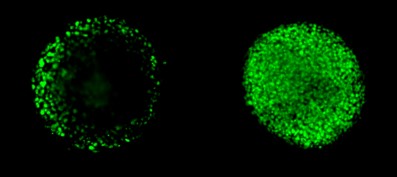
Clearing with Visikol HISTO-M renders the 3D model transparent, allowingfor the detection of every single cell using confocal imaging with a HighContent instrument.

NCI-H2170 spheroids approx 250 um in diameter labeledwith nuclear stain. Left is in PBS and right is the same spheroid afterclearing with Visikol HISTO-M.

3D cellculture in PBS, not cleared. A large majority of cells are not observable.

3D cell culture after clearing withVisikol HISTO-M - every cell in the model can be detected

Number ofcells characterized in before and after clearing NCI-H2170 3D cell culture model
Results are clear withVisikol HISTO-M!
Get the whole picture
Improvedsensitivity and reduction of bias
Significant differences between cleared and non-cleared tumor spheroidshave been observed in the dose response curves of commonantiproliferatives. The application of tissue clearing to 3D cell culturecharacterization has been shown to increase sensitivity to measure doseresponse by an order of magnitude due to the increased number of cells detectedin cleared spheroids. Furthermore, highly significant differences in doseresponse are measured between cleared and non-cleared tumor spheroids due tothe increased level of proliferation in the outermost cells.

NCI-H2170spheroids dosed with cisplatin and evaluated for cell proliferation (Ki67positive cells); A) Dose response relative to vehicle control; B) Absolute cellproliferation score


