visually tracking the progression of disease simultaneous detection of multiple probes drug metabolism and pharmacokinetic studies

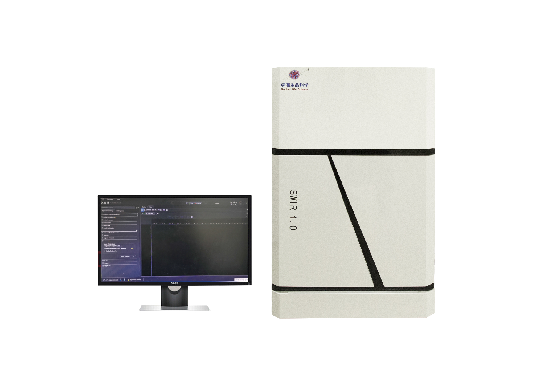
Combining with ultralow background noise and high sensitivity, an advanced gas anesthesia system and a user-friendly operation inter-face etc, SWIR 1.0 can implement both fluorescence intensity imaging and fluorescence lifetime imaging. Through switching the lens, wide-field and zoomed-in imaging modes can be realized in the system. Taking advantage of the ultrafast frame rate, SWIR 1.0 is not only suitable for taking a single picture, but also for taking video. Besides, the system also meets the needs of imaging with long expo-sure time, such as bioluminescence imaging and chemiluminescence imagmg.


tissue autofluorescence in in vivo fluorescence imaging ultralow tissue autofluorescence in near-infrared II region
switchable between wide-field and zoomed-in imaging mode data collection and analysis
compatible with Labview and Matlab
fluorescence intensity imaging collection

Fluorescence intensity imaging interface


lesion areas cannot be distinguised from normal lesion areas can be discriminated by fluorescence
tissue due to enrich-ment of probes in liver lifetime imaging
simple and powerful operation interface step-by-step operation
fluorescence lifetime imaging collection

Fluorescence lifetime imaging interface


gas anesthesia device temperature-control device
In vivo fluorescence intensity and fluorescence lifetime imaging system
Four CCD with cooling temperature at -55°C、-65°C、-85°C and - 190°C can be chosen which can drastically reduce imaging background
Two imaging lens, a 35 mm near-infrared lens and a customized lens, can be adopted to realize wide-field (20 cm x 20 cm) and zoomed-in (2 cm x 2 cm) imaging, respectively.
Combining with the gas anesthesia device and temperature-control device, the small animals can be anesthetized for a long time at 37 °C for in vivo imaging.
Two imaging modules are offered which are capable of fluorescence intensity and fluorescence lifetime imaging.
Near-infrared lasers with various excitation wavelengths are provied to meet the needs of imaging with different probes.

Case studies
FLUORESCENCE-Image guide suregery
Optimal surgery time window of in vivo assembly. (a) comparison of MRI and NIR-II fluorescence imaging (1000 nm long-pass filter) of subcutaneous human ovarian adenocarcinoma. (b) Correction for the tumor size ratio between MRI and NIR-II fluorescence bioimaging method. (c) Optical photo of human ovarian adenocarcinoma peritoneal metastases model (i), the corresponding NIR-II fluorescence bioimaging (1000 nm long-pass filter) results obtained at 22h PI (ii) and the enlargement of the NIR-II nanoprobes labeled large peritoneal metastatic tumors (Nos. 1-8) and ultrasmall lesions (Nos. 9-13) (iii). Scale bar, 1 cm. (d) H&E staining results of tumor margin in Nos.1-8 (scale bars, 0.2 mm) and metastatic lesions in Nos. 9-13 (scale bars, 0.5 mm). Tumors were resected under NIR-II fluorescence bioimaging guidance in (a).
Nat. Commun., 2018, 9, 2898

Case studies
FLUORESCENCE-Vessel imaging
Fluorescence imaging of brain vessels and hintlimb vessels achieved by J-aggregates in varied regions.
J Am. Chem. Soc. 2019, 141,49, 19221-19225
Case studies

FLUORESCENCE-lymphatic imaging
Superior lymphatic imaging with BTC 1070 to ICG and IR26. (a) Digital photograph of a nude mouse. (b) Schematic illustration of the anatomical structure of lymphatic system in the hindlimb of nude mice, green arrow represents the lymphatic drainage from the paw to the sciatic lymph node. (c-g) Fluores-cenc images of lymphatic drainage using IR26, ICG and BTC1070 as contrast agents in the hindlimb of nude mice on an InGaAs camera. Scale bar, 2.5 mm.
Nat. Commun., 2019, 10, 1058

Case studies
FLUORESCENCE-multiplexed imaging
NIR-II multiplexed imaging. (a) Photograph depicting CT-26 tumor bearing mouse for dual-channel biolmninescence imaging. Scale bar, 10 mm. (b) Dual-channel biolmninescence imaging of tumor tissue (upper left) and vessels (lower left) with NIR-I-BPs (challllel 1) and NIR-II-BPs (channel 2). Lower right: The zoomed-in high-magnification dual-channel imaging (lower right) displays fine structure between CT-26 tumor and vessles. Scale bar, 10 mm.

Case studies
FLUORESCENCE-inflammation detection
The ratiometric images (950LP/1100LP) of livers of mice treated with PBS, APAP, or NAC + APAP following by PNllOO probes over time. Livers with hepatotoxicity shows higher fluorescence signals
Angew. Chem. Int. Ed., 2019, 58(24), 8166-8171

Case studies
FLUORESCENE-drug tracking and monitoring
In vivo protein release and activity evaluation. (a) NIR bioimaging of mice at different times after orally gavaging BSA-NPTAT-loaded microcarriers under 730 nm or 808 nm excitation. (d) NIR bioimaging of mice at different times after orally gavaging PEP-NPTAT-loaded microcarriers under 730 nm or 808 nm excitation. (b,e) Corresponding signal intensity curves of (a,d) respectively. (c) In vivo release percentages (Q(t)) of BSA-NPTAT from microcarrier. Mean土 s.d. for n=3.
Nat. Commun., 2017, 8, 14702

Case studies
FLUORESCENCE-Inflammation monitoring
(a) The in vivo bioimaging experimental setup. (b) Photograph of a mouse treated with the microneedle patch. Upconversion luminescent images at (c) 980 and (d) 1180 nm and (e) ratiometric (I980/I1180) channels of microneedle patches taken at different time after lipopolysaccharide-induced inflammation. (f)Ratiometric fluorescence (I980/I1180) of the microneedle patches and corresponding H2O2concentration at different time.
Angew. Chem. Int. Ed., 2018, 57(25), 7518-7522
 Case studies
FLUORESCENCE LIFETIME-In VIVO multiplexed imaging
Lifetime-resolved imaging for IVM of tumour biomarkers. (a) Schematics illustrating animal experiment procedures. (b) Lifetime-resolved images for the MCF-7 and BT-474 tumours are decomposed into the three lifetime channels, repre-sented by the red, green and blue monochromatic image sets. Biomarker expression patterns are calculated by integrating the intensities of each components, nom汕zed by the total intensity over the entire tumour area. (c,d) In vitro western blot (c) and ex vivo immunohistochemistry assay (d) results and calculated biomarker expression patterns of the two tumour subtypes. IVM: in vivo multiplexing; WB: western blot; IHC: immunohistochemistry.
Nat. Nanotechnol., 2018, 13(10), 941-946

Case studies
FLUORESCENCE LIFETIME-Information storage and anti-counterfeiting
(b) Illustration of the time-gated fluorescence imaging system with the magnifying function. (c) Schematics of synchronization and signal collection for lifetime measurement. It is the delay time before the NIR CCD captures the next image. (d) In vivo downshifting (1525 nm) image in bright field. (e) Steady-state fluorescence imaging and (f) time-gated fluorescence imaging with delay time of 1 ms of NaErF4@NaYF4:10%Tm3+@NaYF4 nanocrystals upon pulsed 1208 nm laser excitation. (g) The normalized fluorescence intensity profiles across the (e) black and (f) blue lines of interest. Time-gated fluorescence imag-ing with delay time of (h) 0 ms and (i) 6 ms of two spatially overlapped QR codes with Er-tl and Er-tlO nanocrystals upon 1208 nm excitation. (j) Decoding information of image (h).
Angew. Chem. Int. Ed., 2019, 58(30), 10153-10157

Inorganic fluorescent probes
Near-infrared II lanthanide downshifting nanparticles
The morphology, size and core-shell structure can be precisely controlled. Through doping varied lanthanide ions, exciation (730 nm、808nm、 980nm、1064nm、1208nm、1525 nm) and emission (1060 nm、1155 nm、1289nm、1475nm、1525nm) wavelengths of the lanthanide nanpar-tides can also be regulated. In addition, a tunable lifetime range spanning three orders of magnitude with a single emission band can also be achieved in these nanoparticles (microsecond to milisecond).
Organic fluorescent probes

These fluorescent dyes are stable, wavelength-tunable in the NIR-II region and can be adopted for in-vivo high-contrast bioimaging and multiplexed biosensing

these fluorescent dyes have a tunable emission range from 930 nm to 1070 nm. Due to their super anti-quenching property, these dyes are suitable for vessels and lymphatic imaging as well as in vivofluorescence detection in acid environment.

These fluorescent dyes have a tunable emission wavelength range from 1003 nm to 1118 nm. With good water solubility and easy modification, these dyes are suitable for vessels imaging and the respiratory rate quanification for the awake and anaesthetized animal.

The FD dye has good water solubility, with both excitation (1064 nm) and emission (1080 nm) in the NIR-11 region. After combining with fetal bovine serum (FBS), its intensities can be largely enhanced. The dye is suitable for vessels imaging and the respiratory rate quanification
SWIR 1.0
IMAGING MODALITY
Imaging area : 2 cm * 2 cm ~ 20 cm*20 cm
fluorescence intensity imaging
picture or video with high signal-to noise ratio
fluorescence lifetime imaging
fluorescence lifetime range : 40 μs - 20 ms
fluorescence lifetime resolution : < 5%
CAMERA & OPTICS
Four types of CCD provided
Model | NIR vana-LN | NIR vana | NIR vana-ST | NIR vana-HS |
| Array resolution | 640 X 512
|
| Pixel pitch | 20 um X 20 um |
| Peak Response range (>75%) | 1300nm-1550nm | 900nm - 1620 nm | 900nm - 1700 nm |
| System Noise | 15 e-
| ≤120 e- | <60 e- |
| Cooling Tempature(℃) | -190 | -85
| -65 | -55 |
| Frame Rates(fps) | 2.77/1.75 | 110(@10MHz),55(@5MHz),22(@2MHz)
| 250 |
| Scan Rate | 250KHz , 125 KHz | 10MHz,,5MHz,2MHz |
|
| Max exposure time | > 60min | > 1min | <30sec | <2 us to 1 min |
ANIMAL MANAGEMENT
SERGIVET gas anesthesia device
precise temperature control
ILLUMINATION & FILTERS
various near-infrared lasers
common short-and long-pass near-infrared filters
special laser and filter can be provided upon request
HARDWARE CAPABILITIES
fully automatic system
controlled lens focus
automatic exposure
automactic signal acquisition
automatic fluorescence lifetime calculation
motorized precision translation stage