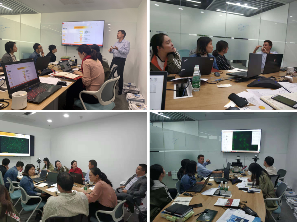
Oct. 23rd , 2018, Dr. Chris Shumate from Etaluma, US
had visited Nuohai Life Science. Lumascope from Etaluma can be widely used in
various live cell, cell spheroid formation, tissue, slides, lab on chip,
bacterial and organisms imaging.
Dr. Shumate had introduced the theory of microscope imaging and
compared the advantage of lumascope with other traditional microscopes. Dr.
Shumate had also discussed deeply with Nuohai team about the marketing,
customers’ demands in China. During the training, Dr. Shumate gave a
demonstration of the lumascope operation and analysis.


About Lumascope
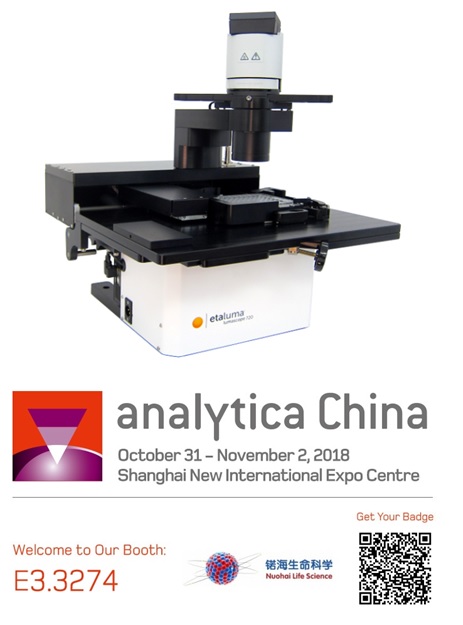
Lumascope, from etaluma US, can be placedinside of the incubator for long term live cell imaging and ensure a stableenvironment for cell growth. It can also be used for small animal, plants andorganisms imaging. As the size of Lumascope is small, it could be take awayeasily and leave in the hood. The opening platform makes it be able to combinewith other instrument or platform. Its light path is simpler and shorter thantradition microscopes, this give the lumascope higher sensitivity and higherimage quality, which is comparable with con-focal microscope. 3-colourfluorescence is available on the microscope (red, green and blue), obejectiverange between 1.25x to 100x, Z-stake imaging is also provided. Culture dish,flask, slides and microplates (up to 1536 well plate) are compatible.Lumaquant, a image analysis software, is specially designed for lumascope. Lumaquantprovides a powerful means to analyze 2D and 2D + Time datasets acquired influorescence, phase contrast, and brightfield microscopy. The analysis recipesin Lumaquant apply state-of-the-art image processing algorithms to enhance,detect and track objects (cells, nuclei, particles, etc.).


Case study

DNA, alpha-tublin, & F-actin in BPAEcells
LS620 image of BPAE cells showing DNA(blue), alpha-tublin (green), & F-actin (red); Olympus 40x objective;LifeTech FluoCell slide #2

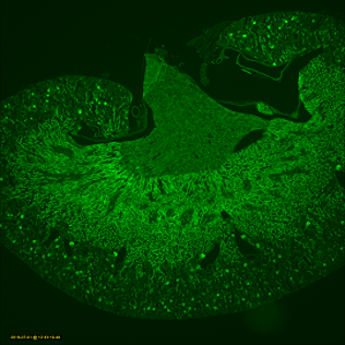
Mouse kidney
Spheroid Formation
FluoVolt Membrane Depolarization in iPSC Cardiac Myocytes
Phase Contrast Time Lapse


