“Simplicity is the ultimate sophistication”
Leonardo Da Vinci
Imaging live cells over longer periods of time, to understand cellular behaviour and dynamic processes, is one of the biggest challenges facing researchers.
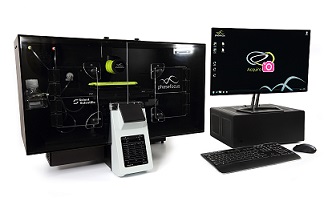
With LivecyteTM, the simple stepwise process means sophisticated long-term time lapse experiments can be set up in under 10 mins.
LessTime: Less Money: More Data
No extensive training requirements
Livecyte's intuitive user interface is designed for ease of use, providing immediate access to the system without the need for costly and time consuming training.
Complete and efficient workflows
Users are guided through each stage of the experiment. The simple Design-Acquire-Analyse functions direct each stage of the experiment, from well selection toimage capture, reducing unproductive laboratory time.
No dedicated consumables
Livecyte works with your standard laboratory consumables, requiring no specialist plates, labels or preparatory kits. Simply seed the cells, place on the microscope and start capturing high contrast, high resolution images.
96 well compatibility
Multiple parameters can be assessed and compared in a single experiment, for more efficient use of laboratory resources.
Automated processing & analysis
Livecyte's Cell Analysis Toolbox software automatically generates a comprehensive rangeof outputs providing multi-parametric data to individual cell level, for more reliable and reproducible cell profiling.
Livecyte - It JustWorks!
The simple solution for sophisticated label-free live cell imaging
See an example of the simple experimental set-up workflow in the short video below.
????Click here to learn more about Livecyte
????

Unlike traditional phase contrast imaging techniques, where images can be corrupted by optical artefacts, Livecyte utilises ptychography, a QPI (Quantitative Phase Imaging) technique, to generate high contrast, information rich images. This innovative new technology permits the quantitative measurement of phenotypic characteristics and dynamic activity for prolonged periods, permitting multiple cell divisions to be monitored.
Livecyte generates a continuous large field of view independent of imaging objective, eliminating the need for image “stitching”. As a result, morphological parameters can be reliably determined providing precise and quantifiable metrics. Furthermore a minimal number of cells move beyond the FOV during imaging, allowing even highly motile cells to be accurately tracked over long periods of time.
The system’s low powered laser illumination reduces the risk of phototoxicity and/or photo-induced behaviour, ensuring cells remain healthy and viable,making it compatible with sensitive cell types, including primary and stemcells.
This ability to analyse cells in a more natural environment provides reassurance that differences in the behaviour of cell populations are genuine rather than a consequence of the imaging technique. validating its suitability for a widerange of applications.
Livecyte also supports widefield fluorescence, permitting labelled and non-labelled protocols to be combined. Supplementing label free images with periodic tracking of fluorescently labelled components keeps phototoxicity to a minimum,allowing additional information to be extracted and results correlated for confident data analysis.
A further strength of the system lies in its capacity to segment individual cells within complex heterogenous cell populations, enabling multiple parameters tobe extracted from a single experiment.
The system comes complete with an extensive array of analysis tools, in corporating both common predefined parameters and customisable options, providing the versatility to tailor assays as required. With its simple, intuitive workflows and interactive graphical tools, users can rapidly design their experiment, setup their acquisition parameters and analyse their data, all with minimal training.
Livecyte includes 12TB data storage for simple straight forward data handling, with data exported as graphs, tables images or video. In addition, design, set up and data analysis can all be completed remotely, providing a flexible and efficient resource for multi-user sites.
This comprehensive system has been specifically designed to create an optimal environment for long term live cell assays, heralding a new era in quantitative phase imaging and providing a gateway to the better understanding of cellular processes .
Common applications include but not limited to:


