Webinar - Livecyte Cell Analysis System by Phasefocus
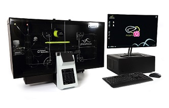
Livecyte is a unique quantitative label free cell analysis system which delivers continuous tracking and analysis of individual cells within large populations, enabling users to gain a more realistic and comprehensive understanding of cell behaviour.
In this webinar, the Phasefocus from UK and researchers from University of Hull (Univerisity of Hull) introduced their study on wound healing of primary fibroblast cells with Livecyte. They analyzed the wound area change and themigration of individual cells. As well as the migration speed, directions, etc., These leads to give more convincing conclusions.
About the presenter

Dr. Rakesh Suman (Phase Focus Limited)
PhD in Neuro Science,from University of Leeds, UK. One of the technical and application developers of Livecyte, he has years of experience in microscope developing and validation. Expert in lable free Ptychographic technology.

Dr. Andrew O'Brien (Phase Focus Limited)
PhD in Optics, from National University of Ireland Galway, Product Manager of Livecyte in Phase Focus Limited. With more than 10 years of experience in biological imaging research and development, he is a core developer and project leader in many optical imaging products. He has in-depth studies with the imaging directions of cell proliferation, cytotoxicity, migration, wound healing, and mitosis.

Amy Qin, Livecyte Product Manager from Nuohai Life Science
Master of Molecular Medicine, University of Sheffield, UK. Now she is the product manager of Livecyte, in Nuohai Life Sciences. She is familiar with a variety of cell imaging equipments on the market, and have experience in culturing hundreds of cell types. She has worked as a research scientist in big biological CRO in China, and have provided new drug research and development services for big pharma such as Pfizer and Merck.
Click here to review the webinar
About Livecyte

Unlike traditional phase contrast imaging techniques, where images can be corrupted by optical artefacts, Livecyte utilises ptychography, a QPI (Quantitative Phase Imaging) technique, to generate high contrast, information rich images. This innovative new technology permits the quantitative measurement of phenotypic characteristics and dynamic activity for prolonged periods, permitting multiple cell divisions to be monitored.
Livecyte generates a continuous large field of view independent of imaging objective, eliminating the need for image “stitching”. As a result, morphological parameters can be reliably determined providing precise and quantifiable metrics. Furthermore a minimal number of cells move beyond the FOV during imaging, allowing even highly motile cells to be accurately tracked over long periods of time.
The system’s low powered laser illumination reduces the risk of phototoxicity and/or photo-induced behaviour, ensuring cells remain healthy and viable, making it compatible with sensitive cell types, including primary and stem cells.
This ability to analyse cells in a more natural environment provides reassurance that differences in the behaviour of cell populations are genuine rather than a consequence of the imaging technique. validating its suitability for a wide range of applications.
Livecyte also supports widefield fluorescence, permitting labelled and non-labelled protocols to be combined. Supplementing label free images with periodic tracking of fluorescently labelled components keeps phototoxicity to a minimum, allowing additional information to be extracted and results correlated for confident data analysis.
A further strength of the system lies in its capacity to segment individual cells within complex heterogenous cell populations, enabling multiple parameters to be extracted from a single experiment.
The system comes complete with an extensive array of analysis tools, incorporating both common predefined parameters and customisable options, providing the versatility to tailor assays as required. With its simple, intuitive workflows and interactive graphical tools, users can rapidly design their experiment, setup their acquisition parameters and analyse their data, all with minimal training.
Livecyte includes 12TB data storage for simple straightforward data handling, with data exported as graphs, tables images or video. In addition, design, set up and data analysis can all be completed remotely, providing a flexible and efficient resource for multi-user sites.
This comprehensive system has been specifically designed to create an optimal environment for long term live cell assays, heralding a new era in quantitative phase imaging and providing a gateway to the better understanding of cellular processes .
Common applications include but not limited to:


