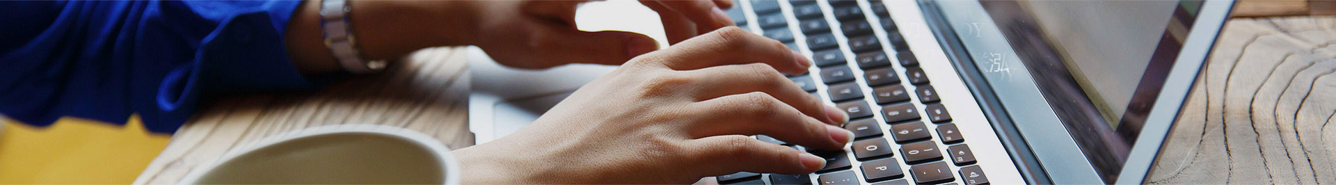
Fig. 1 A ptychographic image of two sheets of molybdenum disulfide, with one rotated by 6.8 degrees with respect to the other. The distances between individual atoms range from a full atomic bond length down to complete overlap.
Image courtesy of Prof. David Muller.
Researchers at Cornell University haveused ptychography to achieve the highest resolution ever produced in anelectron microscope. The team, led by David Muller, Professor of Applied and Engineering Physics, recently reported their findings in the July 19th issue of Nature [1], outlining their ability toresolve images to a resolution of 0.39 A. This level of resolution has enabledthem to visualise individual atoms with greater contrast, and less damage than was previously possible. To put their achievement into perspective, it representsa 2.5x improvement over the conventional imaging performance of their microscope.
Ptychography is a computational imaging technique that utilises sophisticated algorithms to reconstruct animage from overlapping diffraction patterns, created when an object is illuminated. In their paper, Muller’s group use a powerful new electron camera, which they have developed, to record the diffraction patterns with exceptional sensitivity, speed and dynamic range. The world-record resolution was made possible by the combination of detector and ptychography, heralding a new era in electron microscopy.
PhasefocusTM has commercialised ptychography as its proprietary platform technology, creating a portfolio of products to accommodate a wide range of imaging applications, encompassing life science, healthcare, engineering, metrology andmore.
The Phasefocus πbox reconstruction engine, suitable for electron microscopy, is a back-end processing platform that can be connected to a microscope, network or cloud based, to deliver reconstructed ptychographic images to the user’s own software.
Compatible with electrons and any electromagnetic wave, πbox deciphers raw diffraction patterns to streamline image generation, making it applicable across a wide range of imaging modalities including X-ray, Electron microscopy and optical microscopy.
Reference
[1] “Electron ptychography of 2Dmaterials to deep sub-angstrom resolution”, Jiang et. al, Nature, volume 559,pages343–349 (2018) https://doi.org/10.1038/s41586-018-0298-5

About Livecyte
Unlike traditional phase contrast imaging techniques, where images can be corrupted by optical artefacts, Livecyte utilises ptychography, a QPI (Quantitative Phase Imaging) technique, to generate high contrast, information rich images. This innovative new technology permits the quantitative measurement of phenotypic characteristics and dynamic activity for prolonged periods, permitting multiple cell divisions to be monitored.
Livecyte generates a continuous large field of view independent of imaging objective, eliminating the need for image “stitching”. As a result, morphological parameters can be reliably determined providing precise and quantifiable metrics. Furthermore a minimal number of cells move beyond the FOV during imaging, allowing even highly motile cells to be accurately tracked over long periods of time.
The system’s low powered laser illumination reduces the risk of phototoxicity and/or photo-induced behaviour, ensuring cells remain healthy and viable, making it compatible with sensitive cell types, including primary and stem cells.
This ability to analyse cells in a more natural environment provides reassurance that differences in the behaviour of cell populations are genuine rather than a consequence of the imaging technique. validating its suitability for a wide range of applications.
Livecyte also supports widefield fluorescence, permitting labelled and non-labelled protocols to be combined. Supplementing label free images with periodic tracking of fluorescently labelled components keeps phototoxicity to a minimum, allowing additional information to be extracted and results correlated for confident data analysis.
A further strength of the system lies in its capacity to segment individual cells within complex heterogenous cell populations, enabling multiple parameters to be extracted from a single experiment.
The system comes complete with an extensive array of analysis tools, incorporating both common predefined parameters and customisable options, providing the versatility to tailor assays as required. With its simple, intuitive workflows and interactive graphical tools, users can rapidly design their experiment, setup their acquisition parameters and analyse their data, all with minimal training.
Livecyte includes 12TB data storage for simple straightforward data handling, with data exported as graphs, tables images or video. In addition, design, set up and data analysis can all be completed remotely, providing a flexible and efficient resource for multi-user sites.
This comprehensive system has been specifically designed to create an optimal environment for long term live cell assays, heralding a new era in quantitative phase imaging and providing a gateway to the better understanding of cellular processes .
Common applications include but not limited to: