Oct. 25th, 2018, Dr. Chris Shumate, CEO from Etaluma US, visited the School of Pharmacology, Shanghai Jiaotong University, together with Application Engineer and from Nuohai Life Science. Lumascope 720 was showed to the researchers in the school, and Dr. Chris Shumate gave a very detailed introduction about the imaging theory, features and applications of Lumascope 720. After the discussion, Lumascope 720 was placed inside of a traditional incubator and set up a time-lapse cell imaging experiment. This give a better environment for cell growth compared with the live cell workstation. With the extra low toxicity feature, live cell imaging with Lumascope makes experiment easier and cheaper!
Lumascope demonstration is now running country wide, please contact Nuohai Life Science for request a demo!
Contact us from
86-21-37827858
info@nuohailifescience.com

About Lumascope Family

Lumascope, from etaluma US, can be placedinside of the incubator for long term live cell imaging and ensure a stable environment for cell growth. It can also be used for small animal, plants and organisms imaging. As the size of Lumascope is small, it could be take away easily and leave in the hood. The opening platform makes it be able to combine with other instrument or platform. Its light path is simpler and shorter than tradition microscopes, this give the lumascope higher sensitivity and higher image quality, which is comparable with con-focal microscope. 3-colour fluorescence is available on the microscope (red, green and blue), obejective range between 1.25x to 100x, Z-stake imaging is also provided. Culture dish,flask, slides and microplates (up to 1536 well plate) are compatible. Lumaquant, a image analysis software, is specially designed for lumascope. Lumaquant provides a powerful means to analyze 2D and 2D + Time datasets acquired influorescence, phase contrast, and brightfield microscopy. The analysis recipesin Lumaquant apply state-of-the-art image processing algorithms to enhance, detect and track objects (cells, nuclei, particles, etc.).
Case Study
Labon Chip,mimicking the thrombosis
iPSC induced cardiomyocytes beating
Whole blood was stained with Calcein AM to study adhesion of blood derived cells on an endothelial cell layer. Anarterial-like, high pulsatile blood flow profile was applied to induce platelet accumulation. Courtesy Robert Szulcek, LUMC

Taken by LS460, Dog esophagus-134

Taken by LS460, Pine leaf 395

LS560 image of BPAE cells showing alphatubulin; Olympus 10x objective; LifeTech FluoCell slide #2

LS560 image of BPAE cells showing alphatubulin; Olympus 100x (oil) objective; LifeTech FluoCell slide #2
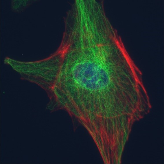
DNA, alpha-tublin, & F-actin in BPAEcells
LS620 image of BPAE cells showing DNA(blue), alpha-tublin (green), & F-actin (red); Meiji 100x (oil) objective;LifeTech FluoCell slide #2.